#Xenon-aided MRIs snap sharper images of lungs
“#Xenon-aided MRIs snap sharper images of lungs”

If you’re a clinician who wants to look inside your patient’s lungs, your options historically have been limited.
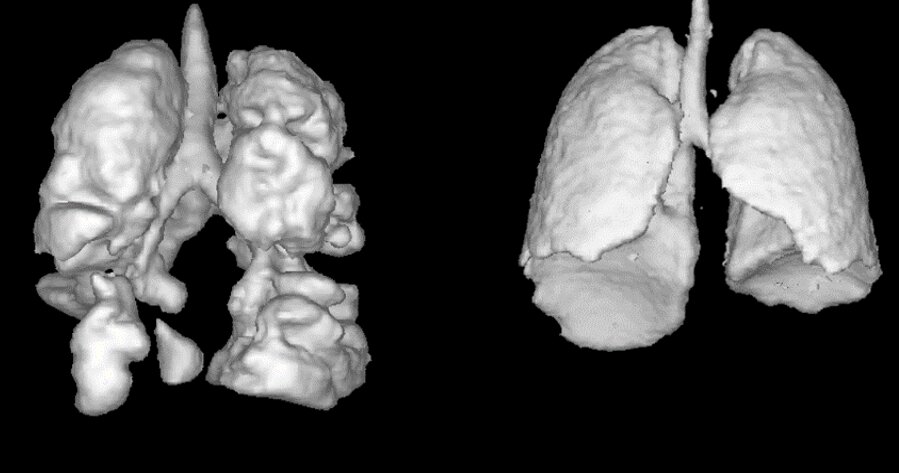
With X-rays or conventional MRI scans, the organs appear mostly as dark cavities in the chest. With CT scans the snapshots are somewhat better, but the low-dose radiation that’s part of the process means CT can’t be used routinely for children.
Lungs—with their mix of water, gas and paper-thin tissue—just don’t image well with conventional MRIs.
Now, professor Alexei Ouriadov of Western’s department of physics and astronomy in the Faculty of Science is establishing a solution that could revolutionize clinicians’ detection of lung ailments and how to treat them.
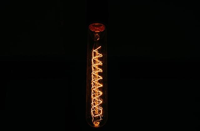
It’s xenon, an inert gas most often used in high-intensity lamps and photo flashes.
When xenon is hyperpolarized and used as a contrast agent in magnetic resonance imaging, the lungs appear lit up like colorful lanterns. Any structural or functional issues are illuminated exponentially better than with ordinary MRIs.
Even better, the scans provide a live look at how the lungs are working, how well they’re transferring life-giving oxygen to red blood cells.
“We get high-resolution information about the lung and we can visualize how it functions,” said Ouriadov, who is leading research to understand the benefits of imaging with xenon.
Patients breathe in a xenon gas blend and hold their breath for 10 seconds while the MRI scanner takes its image. The patient’s body harmlessly absorbs and dissolves the inert xenon in much the same way it handles oxygen, Ouriadov said.
“You can use this technique to observe several lung diseases including asthma, COPD, emphysema, cystic fibrosis, COVID and e-cigarette smoking effects on lung health.”
Interestingly, the patient’s body harmlessly absorbs and dissolves the inert xenon, in much the same way it handles oxygen, so the other organs such as brain, heart, and kidney can be images with xenon. Ouridov said his team is also exploring using xenon to help with brain perfusion imaging.
The U.S. Federal Drug Administration recently approved the use of xenon-129 for imaging in humans, and Ouriadov believes it’s only a matter of time before Health Canada also approves it.
The gas is relatively inexpensive, with a cost of about $20 per dose.
The hitch is that not every facility would have the expertise or equipment to hyperpolarize xenon.
“Once we have approvals, we believe people will be more and more interested in investing into this direction of research,” said Ouriadov. “I think maybe in five years from now, it should be a clinical tool. I am trying to do my very best just to speed up this practice.”
Study confirms longer-term lung damage after COVID-19
Citation:
Xenon-aided MRIs snap sharper images of lungs (2021, June 2)
retrieved 2 June 2021
from https://medicalxpress.com/news/2021-06-xenon-aided-mris-snap-sharper-images.html
This document is subject to copyright. Apart from any fair dealing for the purpose of private study or research, no
part may be reproduced without the written permission. The content is provided for information purposes only.
If you liked the article, do not forget to share it with your friends. Follow us on Google News too, click on the star and choose us from your favorites.
For forums sites go to Forum.BuradaBiliyorum.Com
If you want to read more Like this articles, you can visit our Science category.