#New X-ray technology can improve COVID-19 diagnosis
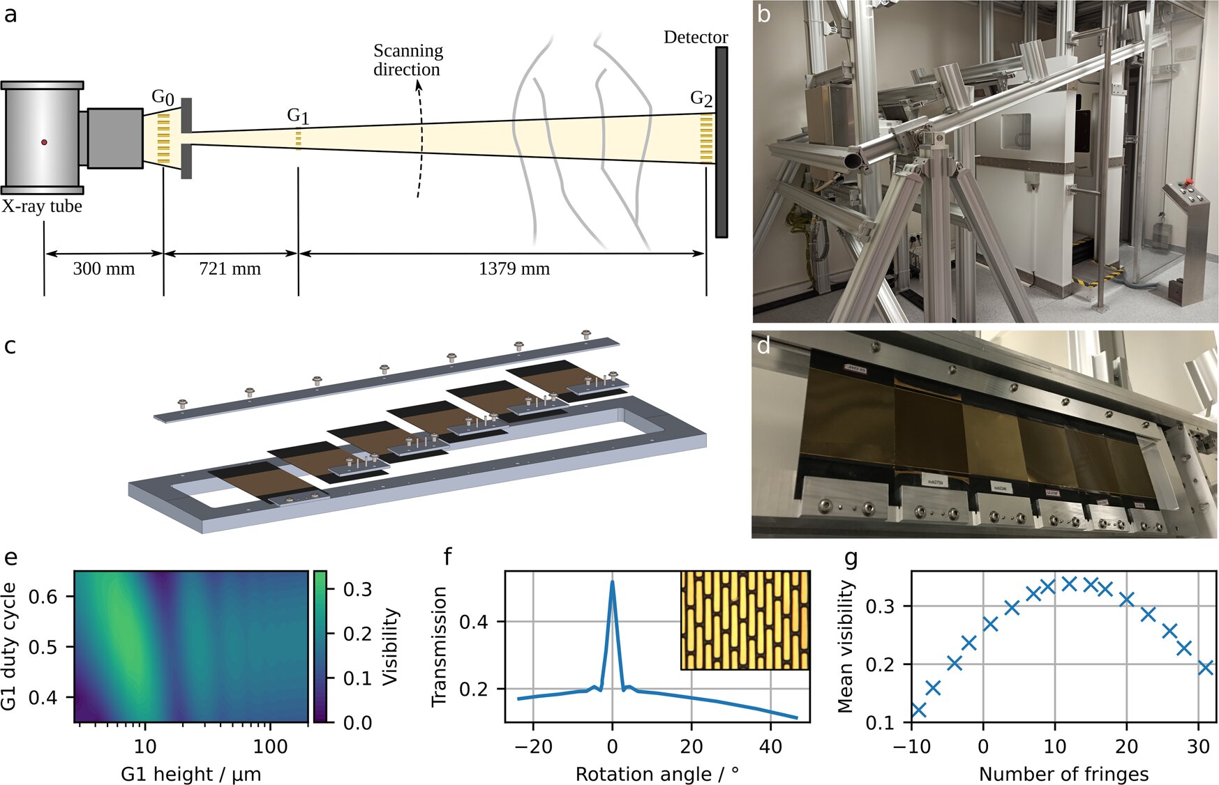
Table of Contents
“New X-ray technology can improve COVID-19 diagnosis”
 for one of the grating structures. <b>g</b> Influence of a number of fringes per frame on mean interferometric visibility. Credit: <i>Communications Medicine</i> (2022). DOI: 10.1038/s43856-022-00215-3")
A research team at the Technical University of Munich (TUM) has, for the first time, produced dark-field X-ray images of patients infected with COVID-19. In contrast to conventional X-ray images, dark-field images visualize the microstructure of the lung tissue, thereby providing additional information. This approach has the potential to provide an alternative to computed tomography (CT), which requires a significantly higher radiation dose.
The lungs of COVID-19 patients are normally visualized using computed tomography (CT). CT technology uses multiple X-ray images from different angles to compute a three-dimensional image. This provides more accurate results than two-dimensional imaging using conventional X-ray technology. The downside, however, is a higher radiation dose due to the large number of X-ray images required.
Dark-field chest X-ray is a new X-ray technology developed by Prof. Franz Pfeiffer. It is paving the road for new possibilities in radiological diagnostics: “During our X-ray examination, we take conventional X-ray images and dark-field images simultaneously. This gives us additional information about the affected lung tissue quickly and easily,” says Franz Pfeiffer, Professor of Biomedical Physics and Director of the Munich Institute of Biomedical Engineering at TUM.
“The resulting radiation dose is fifty times smaller compared to CT machines. Consequently, this method is promising for application scenarios that require repeated examinations over extended periods of time—for example, when investigating the progression of long COVID. This approach might serve as an alternative to computed tomography for imaging lung tissue over prolonged observation periods,” Franz Pfeiffer explains further.
Additional information on lung tissue microstructure
In a new study, published in the journal Communications Medicine, radiologists compared the images of patients with COVID 19 lung disease to those of healthy individuals. They found that distinguishing between sick and healthy individuals was easier using dark-field images than conventional X-ray images. The radiologists were able to differentiate between diseased and healthy lung tissue most readily when both kinds of images—conventional and darkfield—were available.
While conventional X-ray technology relies on the attenuation of X-rays, dark-field X-ray technology utilizes so-called small-angle X-ray scattering. This opens the door to garner additional information on the nature of lung tissue microstructure. Dark-field images can thus provide added value when investigating a variety of lung diseases.
Quantitative evaluation
The research team optimized the X-ray machine prototype to allow them to evaluate the images quantitatively. A healthy lung with many intact alveoli produces a strong dark-field signal and appears bright in the image. In contrast, inflamed lung tissue, with embedded fluid, produces a weaker signal and appears darker in the image. “We normalize the dark-field signal with regard to lung volume to account for lung volume variations between individuals,” explains Manuela Frank, a lead author of the paper.
“Next, we hope to examine further patients. Once we have sufficient dark-field X-ray data available, we intend to use artificial intelligence to support the evaluation process. For conventional images, we have already carried out a pilot project for AI evaluation of our X-ray images,” says Daniela Pfeiffer, professor of radiology and medical director of the study at the TUM University hospital Klinikum rechts der Isar.
Manuela Frank et al, Dark-field chest X-ray imaging for the assessment of COVID-19-pneumonia, Communications Medicine (2022). DOI: 10.1038/s43856-022-00215-3
Citation:
New X-ray technology can improve COVID-19 diagnosis (2022, December 6)
retrieved 6 December 2022
from https://medicalxpress.com/news/2022-12-x-ray-technology-covid-diagnosis.html
This document is subject to copyright. Apart from any fair dealing for the purpose of private study or research, no
part may be reproduced without the written permission. The content is provided for information purposes only.
If you liked the article, do not forget to share it with your friends. Follow us on Google News too, click on the star and choose us from your favorites.
For forums sites go to Forum.BuradaBiliyorum.Com
If you want to read more Like this articles, you can visit our Science category.