#Deep learning for reticular opacity on chest radiographs with interstitial lung disease
“#Deep learning for reticular opacity on chest radiographs with interstitial lung disease”
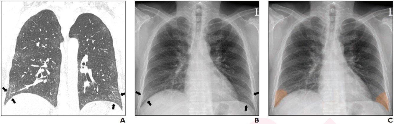
 Coronal chest CT shows bilateral lower lobe pleural-based mild reticular opacities with traction bronchiectasis (arrows). (B) Chest radiograph shows subtle corresponding subpleural opacities in basal lung zone bilaterally (arrows). Only one of six readers detected reticular opacity when interpreting radiograph alone. (C) DLA annotated abnormalities in basal lung bilaterally with probability of 0.99. DLA deemed true-positive for reticular opacity. Five of six readers detected reticular opacity using DLA. Credit: American Roentgen Ray Society (ARRS), <i>American Journal of Roentgenology</i> (<i>AJR</i>)")
According to ARRS’ American Journal of Roentgenology (AJR), radiologists’ sensitivity on chest radiographs for reticular opacity in patients with interstitial lung disease (ILD)—including those with mild disease—improved using a commercially available deep learning algorithm (DLA) from VUNO.
“Use of the DLA improved reader performance and interobserver agreement,” wrote corresponding author Sang Min Lee from Korea’s Asan Medical Center in Seoul, noting that the “benefits were more notable in terms of sensitivity than specificity.”
Lee and team’s retrospective study included 197 patients (130 men, 67 women; mean age, 62.6 years) with surgically proven ILD between January 2017 and December 2018 who underwent preoperative chest radiography and chest CT within a 30-day period. The VUNO Med–Chest X-ray DLA was used to detect lower lobe or subpleural abnormalities; those matching reticular opacity location on CT were deemed true-positives. Six readers (three thoracic radiologists and three residents) independently reviewed radiographs for reticular opacity presence with and without DLA.
In samples from two centers, the VUNO DLA’s accuracy for reticular opacity detection on chest radiograph was 98.5% and 100.0%. Additionally, the DLA improved reader sensitivity from 66.7% to 86.8% in mild, 84.2% to 99.8% in moderate, and 87.3% to 100.0% in severe disease. The DLA also improved interobserver agreement from kappa of 0.517 to 0.870.
“Given the sensitivity of DLA for reticular opacity in mild to moderate disease, the technique could be applied for screening patients with suspected ILD, an area for which chest radiography has historically not performed well,” suggested the authors of this AJR article.
Reduced vs. standard CT dose for lung nodules in children, young adults with cancer
Wooil Kim et al, Utility of a Deep Learning Algorithm for Detection of Reticular Opacity on Chest Radiograph in Patients with Interstitial Lung Disease, American Journal of Roentgenology (2021). DOI: 10.2214/AJR.21.26682
Citation:
Deep learning for reticular opacity on chest radiographs with interstitial lung disease (2021, October 20)
retrieved 20 October 2021
from https://medicalxpress.com/news/2021-10-deep-reticular-opacity-chest-radiographs.html
This document is subject to copyright. Apart from any fair dealing for the purpose of private study or research, no
part may be reproduced without the written permission. The content is provided for information purposes only.
If you liked the article, do not forget to share it with your friends. Follow us on Google News too, click on the star and choose us from your favorites.
For forums sites go to Forum.BuradaBiliyorum.Com
If you want to read more Like this articles, you can visit our Science category.