#Fluorescence imaging based on nanoparticles supports tumor diagnosis
“#Fluorescence imaging based on nanoparticles supports tumor diagnosis”

A malignant tumor (cancer) represents a new organism formed by mutation of normal cells in vivo. It can grow continuously and escape the elimination of the immune system. Therefore, early diagnosis and treatment are essential for the inhibition of tumor growth.
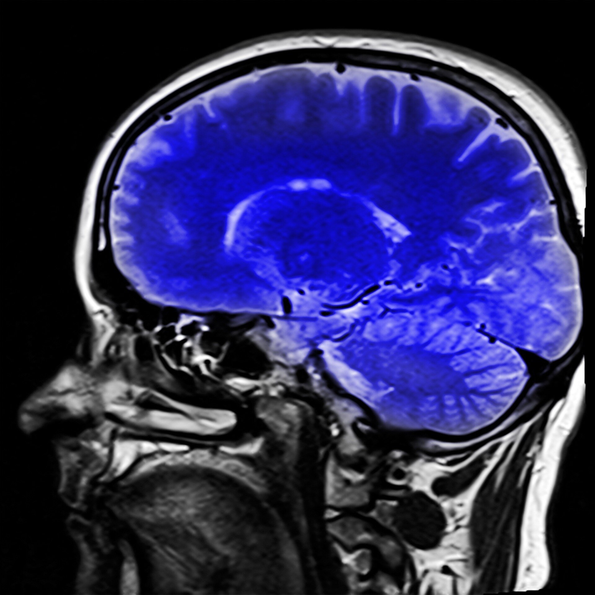
In contrast to conventional means, bioimaging has the potential to accurately locate and diagnose tumors at an early stage by the methods of characterization, visualization and quantification of physiological processes at the cellular and molecular levels. However, the low resolution and dangerous ionizing radiation of CT, PET and SPECT imaging, as well as the high time-consuming of PAI and MRI imaging limit their application in disease treatment.
In a study published in the Journal of Luminescence, a research team led by Prof. Dr. Peng Bo from Xi’an Institute of Optics and Precision Mechanics (XIOPM) of the Chinese Academy of Sciences (CAS) proposed a biocompatible, nontoxic, near-infrared fluorescent tumor targeting nanoparticles (NPs) KHLF(K5HoLi2F10) for the early diagnosis of tumor.
The preparation of the KHLF NPs is according to the atypical hydrothermal method. CH3CH2OK, Ho(NO3)3 ● 6H2O, and LiNO3 were respectively prepared into aqueous solutions of a certain concentration by dissolution in deionized water. After mixing them, PEG was added to the mixture and stirred evenly.
The researchers modified APTES on the novel NPs doped with Ho3+ via a step-by-step method and covalently linked folic acid (FA) to the surface to endow it with the water solubility and tumor targeting. The results of FTIR and zeta potential confirmed the modification process.
The subsequent fluorescence imaging showed that the modification of FA did not significantly weaken the fluorescence intensity of NPs. The biodistribution imaging, in-vivo imaging, in-vitro imaging of various organs, and tumor targeting imaging experiments were carried out. The results showed that this nanoparticle can accurately accumulate on HeLa tumor and generate strong fluorescence signal.
The proposed fluorescence imaging based on nanoparticle has great potential as an efficient biocompatible fluorescent contrast agent for tumor diagnosis.
Q. Fan et al, Tumor imaging of a novel Ho3+-based biocompatible NIR fluorescent fluoride nanoparticle, Journal of Luminescence (2021). DOI: 10.1016/j.jlumin.2021.118007
Citation:
Fluorescence imaging based on nanoparticles supports tumor diagnosis (2021, May 24)
retrieved 24 May 2021
from https://medicalxpress.com/news/2021-05-fluorescence-imaging-based-nanoparticles-tumor.html
This document is subject to copyright. Apart from any fair dealing for the purpose of private study or research, no
part may be reproduced without the written permission. The content is provided for information purposes only.
If you liked the article, do not forget to share it with your friends. Follow us on Google News too, click on the star and choose us from your favorites.
For forums sites go to Forum.BuradaBiliyorum.Com
If you want to read more Like this articles, you can visit our Science category.