#Evolving the surgical microscope
“#Evolving the surgical microscope”
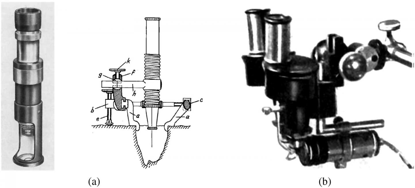
, the modified Brinell microscope (center) and Zeiss binocular microscope adapted by Holmgren (right). Credit: SPIE")
A clear view of anatomical structures is vital for the success of surgery—especially in microsurgery where narrow anatomical cavities or proximity to vulnerable organs and tissues can pose significant risks to patient health. The surgical microscope has evolved to become a powerful tool for improving surgical visualization.
A comprehensive review of surgical microscopes, authored by Baowei Fei, professor of bioengineering and radiology at University of Texas at Dallas, and bioengineering doctoral candidate Ling Ma, relates the evolution of surgical microscopes as they have developed with wider ranges of magnification, longer working distances, better illumination, and more stable supporting structures. Ma and Fei explain how surgical microscopes are modified into slightly different optical configurations and equipped with specific imaging modalities and platforms for different surgical applications.
Beyond the loupe
The first reported surgical microscope in an operating room was used by a young otolaryngology surgeon named Carl Olof Nylén in 1921 at the University of Stockholm. Nylén used a monocular Brinell-Leitz microscope during surgery on a patient with a chronic ear infection. This was a step up from a single lens loupe. The tool was updated a year later by Gunnar Holmgren who developed a binocular microscope for depth perception and an attached light source to accompany the magnification. Holmgren would scarcely recognize today’s surgical microscopes with adjustable magnification, bright and shadow-free illumination, variable working distance to allow manipulation of surgical instruments, and a stable, unobstructed view of the entire operating field.
Though surgical microscopes were not introduced into neurosurgical operating rooms until 1957, when Theodor Kurze at University of Southern California removed a benign tumor from a cranial nerve in a 5-year-old patient, neurosurgery is now the leading market for surgical microscopes.

Versatile visualization, integrated imaging modalities
Surgical microscopy has come a long way. In addition to high-precision optics and flexible mechanical design, versatile visualization features such as 3-D display are now included, as well as integrated imaging modalities. Augmented reality displays, which overlay real-life structures with, say, MRI or OCT images of the structures, improve visualization of the surgical field as well as the multimodal images. Such advanced capabilities are changing clinical practice in the operating room, enabling surgeons to see better and perform challenging procedures with greater ease and success.
Hyperspectral imaging (HSI) and laser speckle contrast imaging (LSCI) modalities are particularly promising since they can be used on-demand anytime during surgery and provide abundant real-time diagnostic data as noncontact and label-free imaging modalities. Ma and Fei note that both HSI and LSCI add very little complexity to the system and take minimal effort for physicians to adopt.
The visualization capabilities and integrated technologies in surgical microscopes continue to expand. With advanced communication technologies and well-developed augmented-reality-assisted platforms, large groups will be able to participate remotely in surgical procedures, sharing a clear view of the surgical field via headsets, smartphones, or large conference room screens.
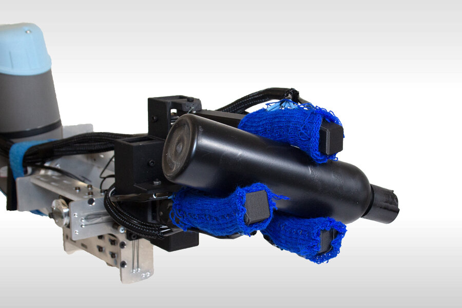
Robotic visualization platforms allow freedom of movement for the surgeon and enable the whole team to observe detailed structures. Integrated technologies, such as an endoscopic micro-inspection tool, can enable the surgeon to ‘bookmark’ a position of the surgical field and visualize the same structure at different angles. Such systems enrich the concept of the surgical microscope with multiple cutting-edge technologies and also provide clear advantages in time, functionality, and ergonomics.
First utilized for otolaryngology, surgical microscopes are contributing to a wide array of microsurgeries, from lymphatic reconstruction to nerve repair. Increasing clinical use of surgical microscopes can be anticipated, and in a greater range of surgical applications.
New microscopy technology augments surgeon’s view for greater accuracy
Ling Ma et al, Comprehensive review of surgical microscopes: technology development and medical applications, Journal of Biomedical Optics (2021). DOI: 10.1117/1.JBO.26.1.010901
Citation:
Evolving the surgical microscope (2021, January 5)
retrieved 5 January 2021
from https://phys.org/news/2021-01-evolving-surgical-microscope.html
This document is subject to copyright. Apart from any fair dealing for the purpose of private study or research, no
part may be reproduced without the written permission. The content is provided for information purposes only.
If you liked the article, do not forget to share it with your friends. Follow us on Google News too, click on the star and choose us from your favorites.
For forums sites go to Forum.BuradaBiliyorum.Com
If you want to read more Like this articles, you can visit our Science category.