#New use of imaging technique could allow early detection of aortic aneurysms
“#New use of imaging technique could allow early detection of aortic aneurysms”

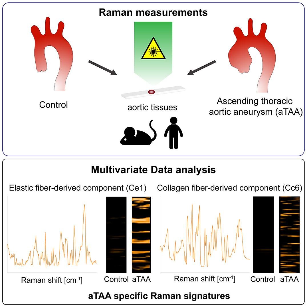
Ascending thoracic aortic aneurisms (aTAAs) occur when the walls of the aorta, the largest blood vessel in the body, weaken and begin to bulge. This can result in rupture or dissection (a tear in the aortic wall), leading to life-threatening bleeding and death. Sometimes these complications can occur before any symptoms of the aneurysm appear. However, an international team led by Hiromi Yanagisawa at the University of Tsukuba and Katja Schenke-Layland at Eberhard Karls University, Tübingen have used Raman microspectroscopy (an analysis technique that uses Raman scattering to probe the structure of atoms and molecules) and Raman imaging to identify signatures in the fibers of the aortic wall that indicate the presence of an aneurysm.
The risk of aTAA is usually assessed by monitoring enlargement of the lumen, the interior space of the aorta. There are no known cardiovascular biomarkers for aortic aneurysms, and changes to the components surrounding the cells (known as the extracellular matrix, or ECM) are not routinely monitored. Changes to the ECM are thought to be involved in the development of aortic aneurysms, because these are commonly seen when there are genetic mutations affecting the ECM. It seems likely that cellular and extracellular (taking place outside of cells) changes to the vessel wall are involved as aneurysms develop.
The research team used both a mouse model of aTAA and human samples to examine the structural and molecular signatures in the aorta. Raman imaging can be used on structures in the body without causing harm and can distinguish different components in tissues by detecting different molecular vibrations. This allowed the team to detect changes to the aortic wall that are characteristic of aneurysms. “Specific elastic fiber-derived components and collagen fiber-derived components were significantly increased in aTAA lesions in both mice and humans,” say co-lead authors Kaori Sugiyama and Julia Marzi. “These human aneurysm-specific marker signatures in the elastic and collagen fibers can be used as biomarkers for aTAA diagnosis.”
These techniques enabled the identification of minor alterations in the ECM structure. This means that this imaging may be used as a diagnostic tool to identify early changes on the aortic wall, known as pre-aneurysmal lesions. Given that, currently, aneurysms can sometimes develop undetected until they cause significant complications, the ability to detect them sooner could save a great many lives.
Other researchers who contributed to this study include Julia Alber and Eva Brauchle of Eberhard Karls University, Masahiro Ando of Waseda University, Yoshito Yamashiro of the University of Tsukuba, and Bhama Ramkhelawon of New York University.
New use of imaging technique could allow early detection of aortic aneurysms
Kaori Sugiyama et al, Raman microspectroscopy and Raman imaging reveal biomarkers specific for thoracic aortic aneurysms, Cell Reports Medicine (2021). DOI: 10.1016/j.xcrm.2021.100261
Citation:
New use of imaging technique could allow early detection of aortic aneurysms (2021, May 24)
retrieved 24 May 2021
from https://medicalxpress.com/news/2021-05-imaging-technique-early-aortic-aneurysms.html
This document is subject to copyright. Apart from any fair dealing for the purpose of private study or research, no
part may be reproduced without the written permission. The content is provided for information purposes only.
If you liked the article, do not forget to share it with your friends. Follow us on Google News too, click on the star and choose us from your favorites.
For forums sites go to Forum.BuradaBiliyorum.Com
If you want to read more Like this articles, you can visit our Science category.