#Nanophotonics enhanced coverslip for phase imaging in biology
“#Nanophotonics enhanced coverslip for phase imaging in biology”
by Light Publishing Center, Changchun Institute of Optics, Fine Mechanics And Physics, CAS </p><div>
<div class="article-gallery lightGallery">
<div data-thumb="https://scx1.b-cdn.net/csz/news/tmb/2021/nanophotonics-enhanced.jpg" data-src="https://scx2.b-cdn.net/gfx/news/2021/nanophotonics-enhanced.jpg" data-sub-html="a The nanophotonics enhanced coverslip (NEC) adds phase imaging capability to a normal microscope coverslip, thereby shrinking bulky phase-imaging methods down to the size of a chip. The less than 200 nm thick design consists of a subwavelength spaced grating on top of an optically thin film, supported by a glass substrate. b Exemplary demonstration of phase-imaging of human cancer cells (HeLa cells) using the NEC. By placing the Petri dish containing the cell culture directly on top of the NEC, pseudo 3D images of the cells are created. The obtained images are similar to those obtained by the conventional phase-imaging technique of differential interference contrast (DIC) microscopy. In the reference image, recorded without the NEC, the cells are mostly invisible. c Use of the NEC device not only enabled visualization of the general shape of the cell, but also features inside of the cell nucleus (left). This was confirmed via comparison with images obtained via conventional DIC microscopy (middle) and fluorescence microscopy (right). Credit: Lukas Wesemann, Jon Rickett, Jingchao Song, Jieqiong Lou, Elizabeth Hinde, Timothy J. Davis, and Ann Roberts">
<figure class="article-img"><img src="https://scx1.b-cdn.net/csz/news/800a/2021/nanophotonics-enhanced.jpg" alt="Nanophotonics enhanced coverslip for phase imaging in biology" title="a The nanophotonics enhanced coverslip (NEC) adds phase imaging capability to a normal microscope coverslip, thereby shrinking bulky phase-imaging methods down to the size of a chip. The less than 200 nm thick design consists of a subwavelength spaced grating on top of an optically thin film, supported by a glass substrate. b Exemplary demonstration of phase-imaging of human cancer cells (HeLa cells) using the NEC. By placing the Petri dish containing the cell culture directly on top of the NEC, pseudo 3D images of the cells are created. The obtained images are similar to those obtained by the conventional phase-imaging technique of differential interference contrast (DIC) microscopy. In the reference image, recorded without the NEC, the cells are mostly invisible. c Use of the NEC device not only enabled visualization of the general shape of the cell, but also features inside of the cell nucleus (left). This was confirmed via comparison with images obtained via conventional DIC microscopy (middle) and fluorescence microscopy (right). Credit: Lukas Wesemann, Jon Rickett, Jingchao Song, Jieqiong Lou, Elizabeth Hinde, Timothy J. Davis, and Ann Roberts" width="720" height="530"/><figcaption class="text-darken text-low-up text-truncate-js text-truncate mt-3">
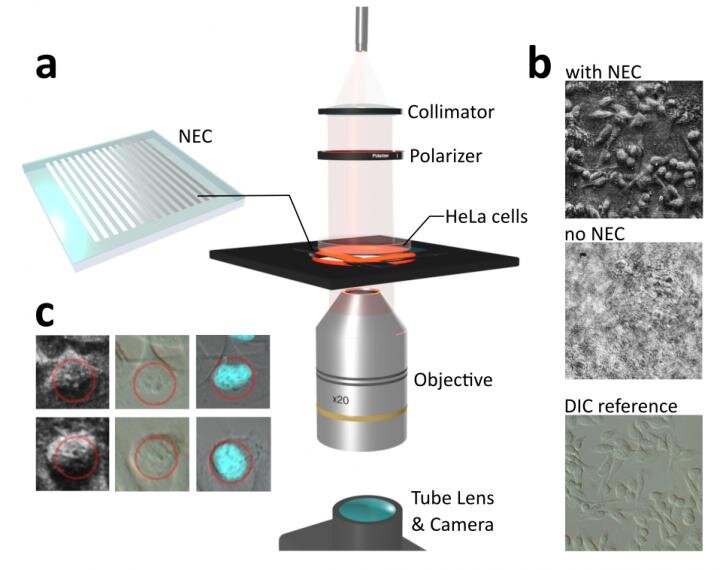
a The nanophotonics enhanced coverslip (NEC) adds phase imaging capability to a normal microscope coverslip, thereby shrinking bulky phase-imaging methods down to the size of a chip. The less than 200 nm thick design consists of a subwavelength spaced grating on top of an optically thin film, supported by a glass substrate. b Exemplary demonstration of phase-imaging of human cancer cells (HeLa cells) using the NEC. By placing the Petri dish containing the cell culture directly on top of the NEC, pseudo 3D images of the cells are created. The obtained images are similar to those obtained by the conventional phase-imaging technique of differential interference contrast (DIC) microscopy. In the reference image, recorded without the NEC, the cells are mostly invisible. c Use of the NEC device not only enabled visualization of the general shape of the cell, but also features inside of the cell nucleus (left). This was confirmed via comparison with images obtained via conventional DIC microscopy (middle) and fluorescence microscopy (right). Credit: Lukas Wesemann, Jon Rickett, Jingchao Song, Jieqiong Lou, Elizabeth Hinde, Timothy J. Davis, and Ann Roberts
</figcaption></figure></div>
The ability to visualize transparent objects such as biological cells is of fundamental importance in biology and medical diagnostics. Conventional approaches to achieve this include phase-contrast microscopy and techniques that rely on chemical staining of biological cells. These techniques, however, rely on expensive and bulky optical components or require changing, and in some cases damaging, the cell by introducing chemical contrast agents. Significant recent advances in nanofabrication technology permit structuring materials on the nanoscale with unprecedented precision. This has given rise to the revolutionary field of meta-optics that aims to develop ultra-compact optical components that replace their bulk-optical counterparts as for example lenses and optical filters. Such meta-optical devices exhibit unusual properties for which they have recently drawn significant scientific interest as novel platforms for imaging applications.
In a new paper published in Light Science & Applications, a team of scientists, led by Professor Ann Roberts from the University of Melbourne node of the Australian Research Council Center of Excellence for Transformative Meta-Optical Systems have developed an ultra-compact, nanostructured microscope coverslip that allows the visualization of unstained biological cells. The device is referred to as a nanophotonics enhanced coverslip (NEC) since it adds phase-imaging capability to a normal microscope coverslip. In their study the researchers demonstrated that by simply placing biological cells on top of the NEC, high-contrast pseudo 3D images of otherwise invisible cells are obtained. The scientists used the example of human cancer cells (HeLa cells) to demonstrate the potential of this new phase-imaging method. The method not only enabled visualization of the general shape of the cancer cells but also made details of the cell nucleus visible. This capacity is crucial since the detection of changes in the structure of biological cells underpins the detection of diseases as for example in the case of malaria.
The version of the NEC presented in the publication differs from a normal coverslip through the addition of a thin-optical film and a nanometer spaced grating. The research team, however, envisage more complex variations of this concept to further extend the capabilities of the method to operation at different wavelengths and integration into highly-specialized optical imaging or microfluidic systems. In conclusion, this research has demonstrated an entirely new phase-imaging method that carries significant potential to be part of future biological imaging systems and mobile medical diagnostic tools.
The scientists summarize the potential of their phase-imaging method: “We designed a nanostructured microscope coverslip that allows us to visualize otherwise transparent biological cells simply by placing them on top of the device and shining light through them. This is an exciting breakthrough in the field of phase-imaging, since our method requires neither the use of bulk-optical components, chemical staining or computational post processing as it is the case with conventional methods.” Prof. Roberts explained.
“The unavailability of medical diagnostic tools in many developing nations is regarded a reason for infectious diseases like malaria and tuberculosis to still be a leading cause of death. Our approach has significant potential to become an inexpensive, ultra-compact phase-imaging tool that could be integrated into smartphone cameras and other mobile devices to make mobile medical diagnostics broadly available.”Dr. Wesemann added.
Holographic histopathology enables fast, precise diagnostics
<hr class="mb-4"/><div class="article-main__more p-4">
<strong>More information:</strong>
Lukas Wesemann et al, Nanophotonics enhanced coverslip for phase imaging in biology, <i>Light: Science & Applications</i> (2021). <a rel="nofollow noopener" target="_blank" data-doi="1" href="http://dx.doi.org/10.1038/s41377-021-00540-7">DOI: 10.1038/s41377-021-00540-7</a>
</div>
<p>
Provided by
Light Publishing Center, Changchun Institute of Optics, Fine Mechanics And Physics, CAS
<!-- print only -->
<div class="d-none d-print-block">
<strong>Citation</strong>:
Nanophotonics enhanced coverslip for phase imaging in biology (2021, May 14)
retrieved 15 May 2021
from https://phys.org/news/2021-05-nanophotonics-coverslip-phase-imaging-biology.html
This document is subject to copyright. Apart from any fair dealing for the purpose of private study or research, no
part may be reproduced without the written permission. The content is provided for information purposes only.
</div>
</div><script id="facebook-jssdk" async="" src="https://connect.facebook.net/en_US/sdk.js"></script>
If you liked the article, do not forget to share it with your friends. Follow us on Google News too, click on the star and choose us from your favorites.
For forums sites go to Forum.BuradaBiliyorum.Com
If you want to read more Like this articles, you can visit our Science category.